NSCLC Pathobiology and Biomarkers

NSCLC is the most common lung cancer in the US and is a heterogeneous disease consisting of 3 major subtypes1,2

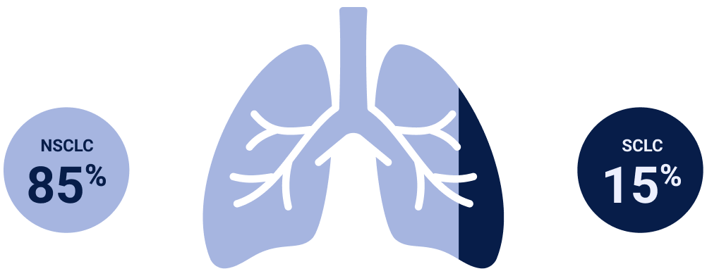
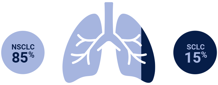
NSCLC=non-small cell lung cancer; SCLC=small-cell lung cancer.

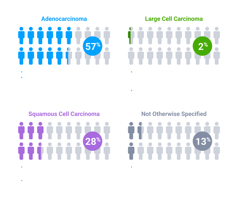
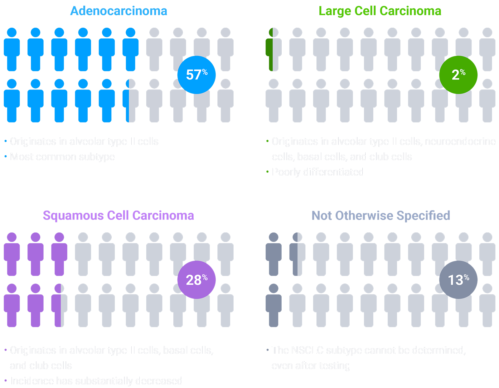
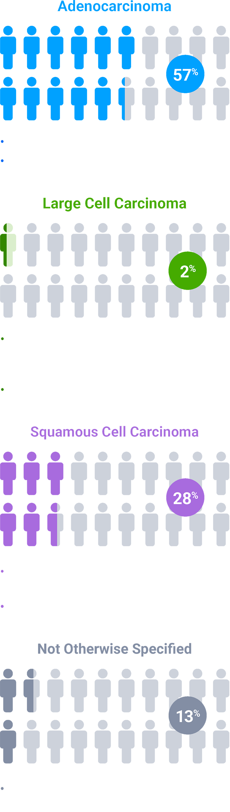
Colors indicate the estimated prevalence of NSCLC subtypes and are for illustrative purposes only. The model does not represent individual patients.

NSCLC=non-small cell lung cancer; SCLC=small cell lung cancer.

Scroll down to learn more Down arrow
Select NSCLC Biomarkers

The molecular basis of lung cancer is complex and develops through a multistep process. Gene alterations and changes in protein expression contribute to the pathogenesis of NSCLC1

Currently, several testing options, such as NGS panels, are able to detect genetic alterations implicated in NSCLC. IHC testing represents a distinct methodology that captures changes in protein expression3

PD-L1 Protein Expression4-​8
~30%

≥1% TPS

High c-Met Protein Overexpression9-11​*
~25%

≥50% TC IHC3+

HER2 Protein Overexpression12,13
~25%

HER2 IHC3+

PD-L1 Protein Expression4-​8
~30%

≥1% TPS

High c-Met Protein Overexpression9-11​*
~25%

≥50% TC IHC3+

HER2 Protein Overexpression12,13
~25%

HER2 IHC3+

This is not an exhaustive list of NSCLC biomarkers. The prevalence of selected biomarkers does not indicate relative efficacy or safety of potential associated therapies. Some biomarkers may have greater characterization across various patient populations.

*Threshold for c-Met protein overexpression is ≥50% tumor cells with strong (3+) staining intensity. EGFR-WT NSQ NSCLC.

EGFR=epidermal growth factor receptor; HER2=human epidermal growth factor receptor 2; IHC=immunohistochemistry; MET=mesenchymal-epithelial transition; NGS=next-generation sequencing; NSCLC=non-small cell lung cancer; NSQ=non-squamous; PD-L1=programmed death-ligand 1; TC=tumor cell; TPS=tumor proportion score; WT=wild type.

Oncogenic Mutations in NSCLC1
Oncogenic mutations in NSCLC graphic
Oncogenic Mutations in NSCLC1
Oncogenic mutations in NSCLC graphic
This is not an exhaustive list of NSCLC biomarkers. The prevalence of selected biomarkers does not indicate relative efficacy or safety of potential associated therapies. Some biomarkers may have greater characterization across various patient populations.

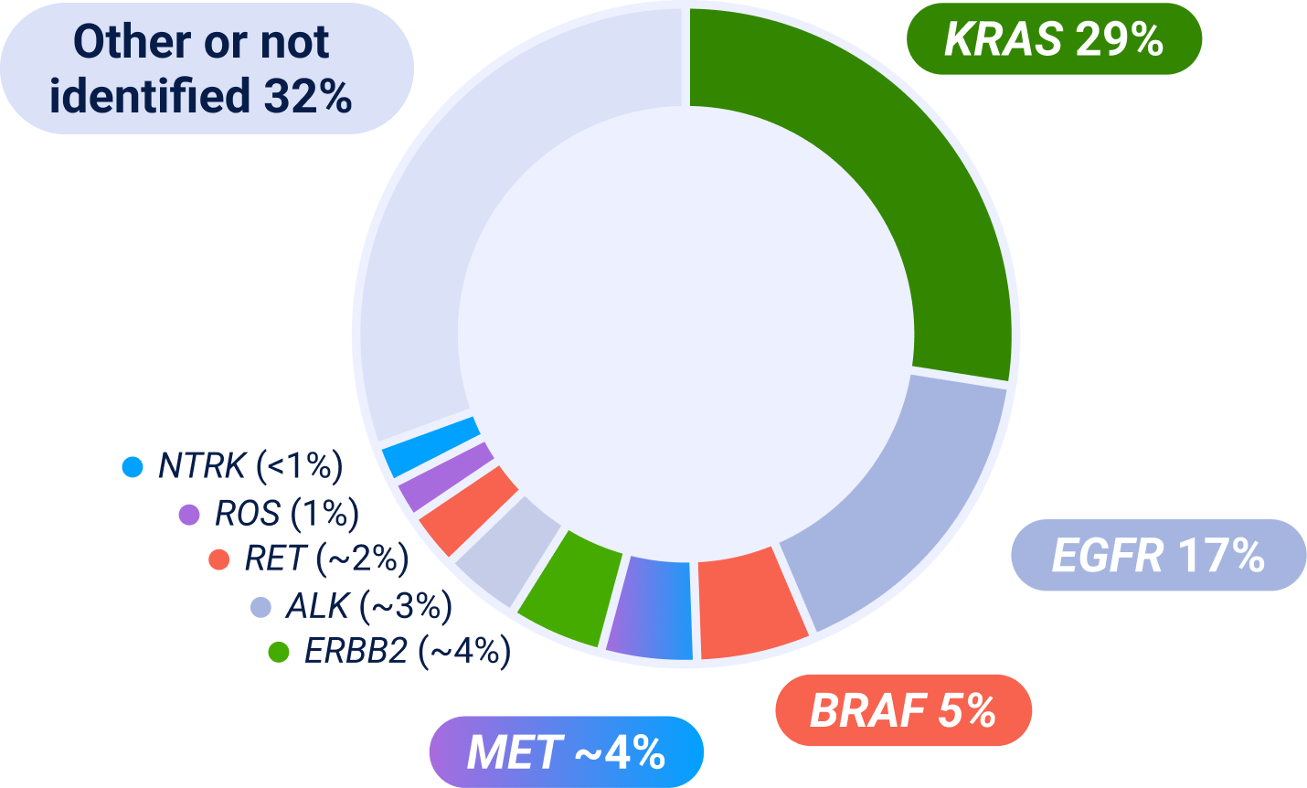
ALK=anaplastic lymphoma kinase; BRAF=B-Raf; EGFR=epidermal growth factor receptor; ERBB2=erb-b2 receptor tyrosine kinase 2; IHC=immunohistochemistry; KRAS=kirsten rat sarcoma viral oncogene homolog; MET=mesenchymal-epithelial transition; NGS=next-generation sequencing; NSCLC=non-small cell lung cancer; NTRK=neurotrophic tyrosine receptor kinase; RET=rearranged during transfection; ROS=ROS proto-oncogene.

References
  1. Thai AA, Solomon BJ, Sequist LV, Gainor JF, Heist RS. Lung cancer. Lancet. 2021;398(10299):535-554. doi:10.1016/S0140-6736(21)00312-3.
  2. Sainz de Aja J, Dost AFM, Kim CF. Alveolar progenitor cells and the origin of lung cancer. J Intern Med. 2021;289:629-635. doi:10.1111/joim.13201.
  3. List of cleared or approved companion diagnostic devices. US Food and Drug Administration. Updated March 5, 2025. Accessed March 31, 2025. https://www.fda.gov/medical-devices/in-vitro-diagnostics/list-cleared-or-approved-companion-diagnostic-devices-in-vitro-and-imaging-tools.
  4. Pawelczyk K, Piotrowska A, Ciesielska U, et al. Role of PD-L1 expression in non-small cell lung cancer and their prognostic significance according to clinicopathological factors and diagnostic markers. Int J Mol Sci. 2019;20(4):824. doi:10.3390/ijms20040824.
  5. Fu F, Deng C, Sun W, et al. Distribution and concordance of PD-L1 expression by routine 22C3 assays in East-Asian patients with non-small cell lung cancer. Respir Res. 2022;23(1):302. doi:10.1186/s12931-022-02201-8.
  6. Aggarwal C, Abreu DR, Felip E, et al. Prevalence of PD-L1 expression in patients with non-small cell lung cancer screened for enrollment in KEYNOTE-001, -010, and -024. Ann Oncol. 2016;27:vi363. doi:10.1093/annonc/mdw378.14.
  7. Dietel M, Savelov N, Salanova R, et al. Real-world prevalence of programmed death ligand 1 expression in locally advanced or metastatic non-small-cell lung cancer: The global, multicenter EXPRESS study. Lung Cancer. 2019;134:174-179. doi:10.1016/j.lungcan.2019.06.012.
  8. Russell PA, Farrall AL, Prabhakaran S, et al. Real-world prevalence of PD-L1 expression in non-small cell lung cancer: An Australia-wide multi-centre retrospective observational study. Pathology. 2023;55(7):922-928. doi:10.1016/j.pathol.2023.08.008A.
  9. Motwani M, Panchabhai S, Bar J, et al. P60.12 Prevalence of c-Met overexpression (c-Met+) and impact of prior lines of treatment on c-Met protein expression in NSCLC. J Thorac Oncol. 2021;16(10):S1169-S1170. doi:10.1016/j.jtho.2021.08.633.
  10. Le X, Aggarwal C, Simmons A, et al. 1303P - METPRO: Evaluating prognostic value of c-Met protein overexpression and concurrent biomarker presence. Ann Oncol. 2024;35(suppl 2):S289. doi:10.1016/j.annonc.2024.08.1360.
  11. Cortot AB, Dubois R, Gregoire V, et al. 165P - Consistency analysis of c-Met protein expression over time in patients with non-squamous non-small cell lung cancer. Ann Oncol. 2024;35(suppl 2):S282. doi:10.1016/j.annonc.2024.08.0173.
  12. Yan M, Schwaederle M, Arguello D, et al. HER2 expression status in diverse cancers: Review of results from 37,992 patients. Cancer Metastasis Rev. 2015;34(1):1557-164. doi:10.1007/s10555-015-9552-6.
  13. Zinner R, Glisson BS, Fossella FV, et al. Trastuzumab in combination with cisplatin and gemcitabine in patients with Her2-overexpressing, untreated, advanced non-small cell lung cancer: Report of a phase II trial and findings regarding optimal identification of patients with Her2-overexpressing disease. Lung Cancer. 2004;44(1):99-110. doi:10.1016/j.lungcan.2003.09.026.

Prevalence estimates are based on multiple sources; survival and prevalence data can vary among studies and datasets because of detection methodology used, patient sample sizes and/or demographics/characteristics. Some patients may have more than one MET aberration and may have overlap with other NSCLC biomarkers.